Studying chalk under a microscope. Common objects under a microscope Various objects under a microscope
Practical work No. 5 Studying chalk under a microscope.
Target: study chalk, sketch its structure, draw conclusions about its origin.
All seas and oceans are inhabited by single-celled organisms whose body is enclosed in a shell. According to modern ideas, they constitute a special type of Foraminifera (from the Latin “foramin” - hole and “ferre” - to carry). Foraminifera shells usually have several chambers with holes in the walls through which the prolegs protrude.
Most foraminifera live at the bottom of the seas, since their heavy shells prevent them from floating to the surface of the water. But there are species that live in the water column; their shells have spines that increase the overall surface, making it easier to float in the aquatic environment
Calcareous shells of dead foraminifera settle to the bottom of the sea. Over time, they are compressed, forming layers of sedimentary rocks - limestone (chalk). Man has long appreciated the merits of sedimentary rocks formed from the skeletons of protozoa. For example, limestone was used in the construction of Egyptian pyramids, temples of Vladimir-Suzdal Rus', snow-white houses of Sevastopol, old buildings in Paris, Rome, Vienna and other cities of the world.
Radiolarians, or rays, are exclusively marine protozoans. Radiolarians inhabit the southern seas with high concentrations of salts. They live mainly in the upper, more oxygenated layers of water.
Radiolarians are characterized by a variety of forms. The most common are spherical radiolarians with long filamentous pseudopods and radially arranged rays of a siliceous skeleton. This is where their second name comes from - rays (see Fig. 8).
A characteristic feature of these protozoa is the presence of an intracellular central capsule and an internal skeleton. Inside the capsule there are one or more nuclei and inclusions of organic substances, such as drops of fat. This makes radiolarians lighter, and they “float” in the water column.
Radiolarians feed on the smallest algae and protozoa, capturing them with their pseudopods.
Like foraminifera, radiolarians play an important role in the formation of sedimentary rocks. Dense layers consisting of radiolarian skeletons are technically called mountain flour or tripoli. It is used for polishing metal and glass products, as well as for making fine sandpaper.
Complete the tasks:
Read the text;
Explain the origin of chalk (limestone) - in writing in a notebook;
What conditions are necessary for the formation of chalk (limestone)?
How people use chalk (limestone) - writing in notebooks;
Draw in your notebook what chalk looks like under a microscope, several radiolarians and foraminifera;
Draw a conclusion about the structure of chalk (limestone) - in writing in a notebook.
A truly powerful microscope is not something you buy for fun, but if you have one, it won’t sit idle. We have proven more than once that even the most ancient trinket in the house becomes an incredible, surreal, amazing, and sometimes even frightening work of art when you look at it through a microscope. It's like a peephole into a parallel world.
Don't you know what I mean? Then take a look at the mind-shocking enlarged images:
8. Chalk
Life-size chalk [publicphoto.org]Chalk is used at school to play hopscotch. If you grind it into powder, it looks like sand and something else... In general, chalk, as we know it, is not very interesting.
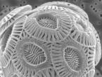
 Close-up: Foraminifera [PLOS Biology]
 Hmm, looks like a soccer ball. In fact, foraminifera shells are the main component of chalk. Foraminifera are the simplest single-celled organisms that have an exoskeleton (shell).
 Kosher salt full size [blogspot.ru]
 Kosher salt is coarser than regular salt and has the property of absorbing the blood of meat, like salt Dracula.
 Close-up of kosher salt [Museum of Science]
 The kosher salt crystal strongly resembles an ancient temple.
 Kosher salt crystals under a microscope [science photo library]
 And here’s another picture - so you can see that all kosher salt consists of “pyramids”.
 Life size orange juice [blogspot.ru]
 Here is the most ordinary orange juice, frankly orange in color, but what will we see under a microscope?
 Orange juice under a microscope [telegraph.co.uk]
 As it turns out, orange juice contains just a hint of orange, more reminiscent of the view inside a kaleidoscope. So now you know that when you enjoy orange juice in the morning, you are drinking liquefied shards in every color of the rainbow.
5. Snow
 Our favorite snow [picturesofwinter.net]
 Extraordinarily beautiful pieces of icy poetry, capable of causing sincere childish joy, as well as falling like an uncontrollable snow storm on an unlucky traveler who had the temerity to find himself outside on a particularly frosty winter day.
 Snow magnified under a microscope [Science Musings]
 Yes, and this is not a child’s paper craft, this is a real snowflake under a microscope. Well, this proves to us once again that nature is imperfect!
 www.wired.com ]
 Let's look at the snow under a microscope again.
4. Anatomy of insects
 Normal size fly [jhunewsletter.com]
 Fly Tsokotukha.
 Close-up of a fly [Wikimedia Commons]
 Looks like a square scorpio!
It is quite possible that after what you have seen, you will no longer carelessly tolerate the proximity of these all sorts of harmful insects.
A tick bite can cause Lyme disease. And here is a picture of what he bites with (scientifically called a hypostome):
 “What a cute tongue you have!”
 This hypostome belongs to the black-eyed tick. Now take a look at the blacklegged tick's knife-shaped mouth:
 Dangerous creature
 And here is an enlarged mosquito sting:
 A mosquito sting under a microscope [Ben133uk]
 This is how they drink our blood. So there is no need to regret the next mosquito killed by your hand.
 Sea water close-up [wordpress.com]
 Water is life.
 Microorganisms found in sea water [N. Sullivan/NOAA/Department of Commerce]
 It is not the water itself, but those who inhabit it. All 247 quadrillion microorganisms. These are diatoms - the general name for dead algae that flood the ocean and, one way or another, sometimes get into your body (when swimming in the sea, for example). Some look delicious. Most, unfortunately, look like either cigars or industrial waste.
 Life Size Fly Ash [www.manatts.com]
 You see fly ash all the time, you just don't know what it is. And this is crushed coal, used to strengthen concrete and asphalt. True, it is very radioactive, so you should not come close to a cloud of such a mixture.
 Fly ash under a microscope [wikimedia.org]
 Under a microscope, fly ash looks like a dead planet with countless craters and lifeless, rocky islands. Or maybe this is just another soap party. Or anything else - depending on your imagination, you can voice your options in the comments.
1. Shark skin
 Normal Shark Skin Size [wordpress.com]
 Sharks are amazing creatures: if a shark stops moving, it dies, a shark can smell a tiny drop of blood in a huge volume of water, unborn shark babies eat each other in the womb until only one remains. The only thing about her that doesn't deserve attention is her skin.
 Shark skin under a microscope [George Lauder]
 Oh, no, her skin, it turns out, is also extremely unusual. It's made of teeth. By the way, they are called denticles, and their purpose is to reduce water resistance when the shark moves.
 Shark skin, enlarged many times [Australian Museum]
 Let's increase it further. Under a microscope, shark skin resembles sharp teeth, which is why it was previously used as a polishing material (in modern times, sandpaper is used). Borazo is the name given to shark skin with polished scales, which is the most expensive leather in the world.
The human body is such a complex and well-coordinated “mechanism” that most of us cannot even imagine! This series of photographs taken using electron microscopy will help you learn a little more about your body and see what we cannot see in our ordinary lives. Welcome to the authorities!
Alveoli of the lungs with two red blood cells (erythrocytes). (photo CMEABG-UCBL/Phanie)

30x enlargement of the base of the nail.

The iris of the eye and adjacent structures. In the lower right corner is the edge of the pupil (blue). (photo by STEVE GSCHMEISSNER/SCIENCE PHOTO LIBRARY)

Red blood cells fall out (so to speak) from the broken capillary.

Nerve ending. This nerve ending was dissected to reveal vesicles (orange and blue) containing chemicals that are used to transmit signals in the nervous system. (photo by TINA CARVALHO)

Clotted blood.

Red blood cells in the artery.

Human lungs.

Taste receptors on the tongue.

Eyelashes, 50x magnification.

Finger pad, 35x magnification. (photo by Richard Kessel)

Sweat pore that comes to the surface of the skin.

Blood vessels coming from the optic nerve nipple (where the optic nerve enters the retina).

The egg that gives rise to a new organism is the largest cell in the human body: its weight is equal to the weight of 600 sperm.

Sperm. Only one sperm penetrates the egg, breaking through the layer of small cells that surround it. As soon as he gets into her, no other sperm can do this.

Human embryo and sperm. The egg was fertilized 5 days ago, and some remaining sperm are still attached to it.

An 8-day embryo at the beginning of its life cycle...
 Without a doubt, the microcosm can impress even those who have decided to connect their lives with science. What can we say about inquisitive beginners or schoolchildren, it surprises even when a person is internally ready for it. And once again this will be proven by studying the chalk under microscope. Laboratory work of the same name is included in the 7th grade biology curriculum. However, it will be much more interesting for young biologists to figure it out on their own, experiment and formulate their first conclusions.
Studying chalk under a microscope It is advisable to carry out at that stage of training when the researcher is able to properly handle an optical instrument - understands what illumination, focusing, etc. are. Quite a lot has been written about this, and I would like to place the main emphasis on the theoretical and practical parts of the experiment.
Being a rock of organic origin, chalk contains the remains of microscopic single-celled organisms. These are primarily radiolarians. They can have a very bizarre shape and often turn out to be different from one another. They are distinguished by the presence of pseudopods - processes that give the body the ability to move. A skeletal design that strongly resembles a twisted shell, reduced many times over, is also common. In addition, foraminifera can be found in the form of whitish shells, mainly consisting of calcium carbonate. Particles of sea or river algae are also mixed into this. This is the unusual composition of a seemingly homogeneous white solid substance, a careful study of which under a microscope will completely change the previously formed idea.
Now about practice. The study of chalk under a microscope should be carried out using the bright field method in transmitted light. This means turning on the lower light (for those models in which it is built-in) or adjusting the mirror (if natural lighting is implemented).
The experiment is carried out in several stages:
- It is necessary to grind the chalk to a powder state.
- The resulting chalk dust is carefully poured onto a glass slide - in a small layer with a mound in the center.
- Using a pipette, drop one drop of water onto the glass with chalk.
- The prepared microspecimen is located strictly under the lens and centered on the table.
- Research begins with the lowest magnification, then the magnification gradually increases.